What are the symptoms of mycosis fungoides?
Mycosis fungoides is a type of cutaneous T-cell lymphoma (CTCL), a cancer that primarily affects the skin. It is characterized by the proliferation of malignant T-lymphocytes in the skin, and its symptoms can vary in presentation. Here are the common symptoms associated with mycosis fungoides:
1. Skin Lesions:
- Plaques: The most typical presentation includes red, raised plaques that may be scaly or itchy. These plaques can often resemble eczema or psoriasis.
- Patches: Early lesions may appear as flat, discolored patches on the skin that are typically less than 1 centimeter in diameter.
- Tumors: As the disease progresses, some patients may develop larger tumors or nodules, which can be ulcerated and may bleed.
- Skin Thickening: Thickened areas of skin (known as lichenification) can develop in response to chronic irritation or scratching.
2. Itching:
- Pruritus: Intense itching is a common symptom and can be particularly bothersome for patients.
3. Changes in Skin Color:
- Hypopigmentation or Hyperpigmentation: Some lesions may lead to lighter (hypopigmented) or darker (hyperpigmented) areas of skin.
4. Erythema:
- Redness of the Skin: Areas of affected skin may appear red or inflamed due to the underlying immune response.
5. Nodal Involvement:
- Lymphadenopathy: In more advanced stages, individuals may have enlarged lymph nodes, indicating that the disease may have spread beyond the skin.
6. Systemic Symptoms:
- In advanced stages of mycosis fungoides, systemic symptoms may appear, including:
- Fever
- Weight Loss
- Night Sweats
- Fatigue
7. Other Symptoms:
- Secondary Infections: Because the skin barrier is compromised, affected individuals may be more prone to skin infections.
Progression:
- Mycosis fungoides typically has a slow progression, and the symptoms can evolve over time. The disease can sometimes mimic other skin conditions, which can lead to delays in diagnosis.
Conclusion:
Mycosis fungoides primarily presents with specific skin lesions, including plaques, patches, and tumors, accompanied by symptoms like itching and erythema. If you or someone you know is experiencing these symptoms, particularly persistent or worsening skin changes, it is crucial to seek evaluation by a healthcare professional, such as a dermatologist or oncologist, for appropriate diagnosis and management. Early detection and treatment can lead to better outcomes for patients with this condition.
What are the causes of mycosis fungoides?
The exact causes of mycosis fungoides, a type of cutaneous T-cell lymphoma (CTCL), are not fully understood; however, several factors and hypotheses are believed to contribute to the development of the disease. Here are some of the potential causes and risk factors associated with mycosis fungoides:
1. Genetic Factors:
- Chromosomal Abnormalities: Certain chromosomal abnormalities and genetic mutations have been associated with T-cell lymphomas, including mycosis fungoides. These genetic changes may affect the regulation of the immune system and cell growth.
2. Immune System Dysfunction:
- Immune Response: An abnormal response of T-lymphocytes (a type of white blood cell) may play a role in the pathogenesis of mycosis fungoides. The malignant T-cells proliferate in the skin, leading to the characteristic lesions.
3. Environmental Factors:
- Chemical Exposures: Some studies suggest that exposure to certain chemicals (e.g., those found in agricultural pesticides, solvents, or heavy metals) may be associated with an increased risk of developing mycosis fungoides.
- Radiation Exposure: Prior exposure to ionizing radiation such as radiation therapy has been mentioned as a potential risk factor for developing CTCL.
4. Viral Infections:
- Human Immunodeficiency Virus (HIV): There is an increased risk of various lymphomas, including mycosis fungoides, among individuals with HIV/AIDS due to compromised immune function.
- Other Viruses: Some studies have explored the role of other viral infections, such as human herpesvirus 6 (HHV-6) and Epstein-Barr virus (EBV), in the development of mycosis fungoides, but the evidence remains inconclusive.
5. Chronic Skin Conditions:
- Pre-existing Dermatological Disorders: People with chronic inflammatory skin conditions (such as psoriasis or eczema) may have an altered immune response that could predispose them to the development of mycosis fungoides.
6. Age and Gender:
- Demographics: Mycosis fungoides is more common in adults, particularly those over the age of 50, and has a slight male predominance, indicating that age and gender might influence susceptibility.
7. Family History:
- Genetic Predisposition: A family history of lymphoproliferative disorders may increase the likelihood of developing mycosis fungoides, suggesting a potential inherited component.
Conclusion:
While the precise cause of mycosis fungoides remains poorly understood, a combination of genetic, environmental, immune system factors, and potential viral influences are believed to contribute to its development. Further research is needed to clarify the interplay of these factors and improve our understanding of this condition. If you have concerns about mycosis fungoides, particularly with regard to symptoms or risk factors, it is advisable to consult a healthcare professional for evaluation and guidance.
How is the diagnosis of mycosis fungoides made?
The diagnosis of mycosis fungoides, a type of cutaneous T-cell lymphoma (CTCL), involves a comprehensive evaluation, including clinical assessment, laboratory tests, and imaging studies. Here’s a structured approach to how the diagnosis is typically made:
1. Clinical Evaluation:
- Medical History: A thorough history is taken to understand the onset and progression of symptoms. The patient is asked about skin changes, itching, duration of lesions, and any associated systemic symptoms (e.g., fever, weight loss).
- Physical Examination: A detailed skin examination is conducted to identify characteristic lesions, such as patches, plaques, or tumors. The doctor will also check for lymphadenopathy (enlarged lymph nodes) and any systemic signs.
2. Dermatological Assessment:
- The specific appearance of lesions is crucial in diagnosing mycosis fungoides. The disease may exhibit different stages, and the morphology of lesions can guide the clinician.
3. Skin Biopsy:
- Histopathological Examination: A definitive diagnosis of mycosis fungoides is usually made with a skin biopsy. A sample of affected skin is taken and examined microscopically for:
- Atypical T-lymphocytes (cancerous cells) in the epidermis and dermis.
- Characteristic findings, such as “cerebriform” nuclei in the lymphocytes.
- Immunohistochemistry: Special staining techniques may be used on the biopsy samples to identify the presence of specific T-cell markers. This helps differentiate mycosis fungoides from other skin conditions.
4. Blood Tests:
- Peripheral Blood Smear: In some cases, a blood sample may be examined to check for abnormal T-cells or other blood abnormalities, but it is not typically used for the initial diagnosis.
5. Imaging Studies:
- Radiological Examination: If the disease is suspected to have spread beyond the skin, imaging studies such as CT scans or PET scans may be performed to assess lymph node involvement or detect any systemic spread of the disease.
6. Staging:
- If diagnosed, staging will be evaluated to determine the extent of the disease. This classification will guide treatment plans. Staging considers the skin lesions’ size and location, lymph node involvement, and any systemic symptoms.
7. Differential Diagnosis:
- Given that mycosis fungoides can mimic other skin conditions (e.g., eczema, psoriasis, or other skin lymphomas), a careful differential diagnosis is crucial to rule out these conditions.
Conclusion:
The diagnosis of mycosis fungoides is primarily established through clinical evaluation, biopsy, and histopathological analysis. If you suspect you or someone you know may have mycosis fungoides, it is important to seek evaluation from a dermatologist or oncologist with experience in managing skin lymphomas. Early diagnosis and intervention can improve outcomes and quality of life.
What is the treatment for mycosis fungoides?
The treatment for mycosis fungoides, a type of cutaneous T-cell lymphoma, depends on several factors, including the stage of the disease, the extent of skin involvement, the presence of symptoms, and the patient’s overall health. The primary goal of treatment is to manage symptoms and control the progression of the disease. Here are the common treatment options:
1. Topical Treatments:
- Corticosteroids: Potent topical steroids may be used to reduce inflammation and control symptoms related to skin lesions. These are often the first line of treatment for early-stage disease.
- Topical Chemotherapy: Agents like chlormethine (Mustargen) or bexarotene can be applied directly to the skin to help reduce tumor burdens.
- Tazarotene: This topical retinoid can also help in managing skin lesions.
2. Phototherapy:
- Narrowband UVB: This form of light therapy can be effective for treating mycosis fungoides, especially in early stages or with localized skin involvement.
- PUVA (Psoralen + UVA): This treatment combines a photosensitizing agent (psoralen) with UVA light to target lymphoma cells in the skin.
3. Systemic Treatments:
For more advanced stages of mycosis fungoides that are not responsive to topical therapies, systemic treatments may be necessary:
- Chemotherapy: Traditional chemotherapy agents such as chlorambucil, combination chemotherapy, or gemcitabine may be used, particularly for advanced or aggressive disease.
- Targeted Therapies:
- Bexarotene: An oral retinoid that is effective for treating advanced stages.
- Brentuximab vedotin: An antibody-drug conjugate that targets CD30+ cells, which may be helpful in some cases.
- Immunotherapy:
- Interferon: This can help boost the immune response against cancerous cells.
- Pembrolizumab or Nivolumab: These immune checkpoint inhibitors may be used in selected patients with refractory disease.
4. Radiation Therapy:
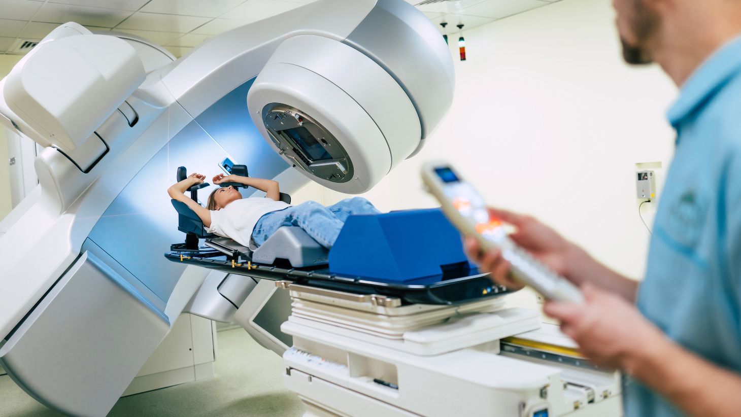
- Localized Radiation: Radiation can be effective for treating skin lesions that are localized, as well as lymph nodes, especially in advanced stages.
- Total Skin Irradiation: In cases of generalized skin involvement, this treatment option targets the entire skin surface and may be used for patients with more widespread disease.
5. Stem Cell Transplantation:
- In cases of advanced mycosis fungoides that have not responded to other therapies, stem cell transplantation may be considered, particularly if there is a matched donor available.
6. Supportive Care:
- Symptom Management: Managing symptoms such as itching and discomfort is crucial. This may include antihistamines for itching and moisturizers for dry skin.
- Psychosocial Support: Counseling, support groups, and educational resources can help patients cope with the emotional aspects of living with a chronic condition.
Conclusion:
The treatment of mycosis fungoides is tailored to the individual based on the stage of the disease and associated symptoms. Management may involve a combination of topical agents, phototherapy, systemic treatments, radiation therapy, and supportive care. Early diagnosis and treatment are vital for improving outcomes. If you or someone you know is dealing with mycosis fungoides, it’s important to collaborate with a healthcare provider or a specialist in dermatology or oncology who is knowledgeable about this condition for optimal care.
What is the survival rate for mycosis fungoides?
Mycosis fungoides, the most common form of cutaneous T-cell lymphoma (CTCL), has a variable survival rate based on factors such as the stage at diagnosis, age, general health, and response to treatment. Here’s a general breakdown:
- Early Stages (IA, IB, and IIA):
- People diagnosed in the early stages often have a favorable prognosis. The 5-year survival rate is estimated at around 85-90%.
- Early-stage patients may live for many years, sometimes with only mild symptoms that can be managed with skin-directed treatments (like phototherapy or topical steroids)【31†source】.
- Advanced Stages (IIB, III, IV):
- As the disease progresses to more advanced stages, it can spread beyond the skin to lymph nodes or internal organs, reducing survival rates.
- For stage IIB, the 5-year survival rate is estimated at 65-70%.
- In stages III and IV, the 5-year survival rate generally decreases to around 20-50%, with stage IV cases typically experiencing the most aggressive progression【31†source】 .
- Impact of Age and Overall Health:
- Older age and the presence of other health issues can lower survival rates in mycosis fungoides, particularly in advanced stages.
Treatment for mycosis fungoides is often long-term and personalized, aiming to manage symptoms and improve quality of life rather than cure.

Leave a Reply
You must be logged in to post a comment.