What are the symptoms of periventricular leukomalacia?
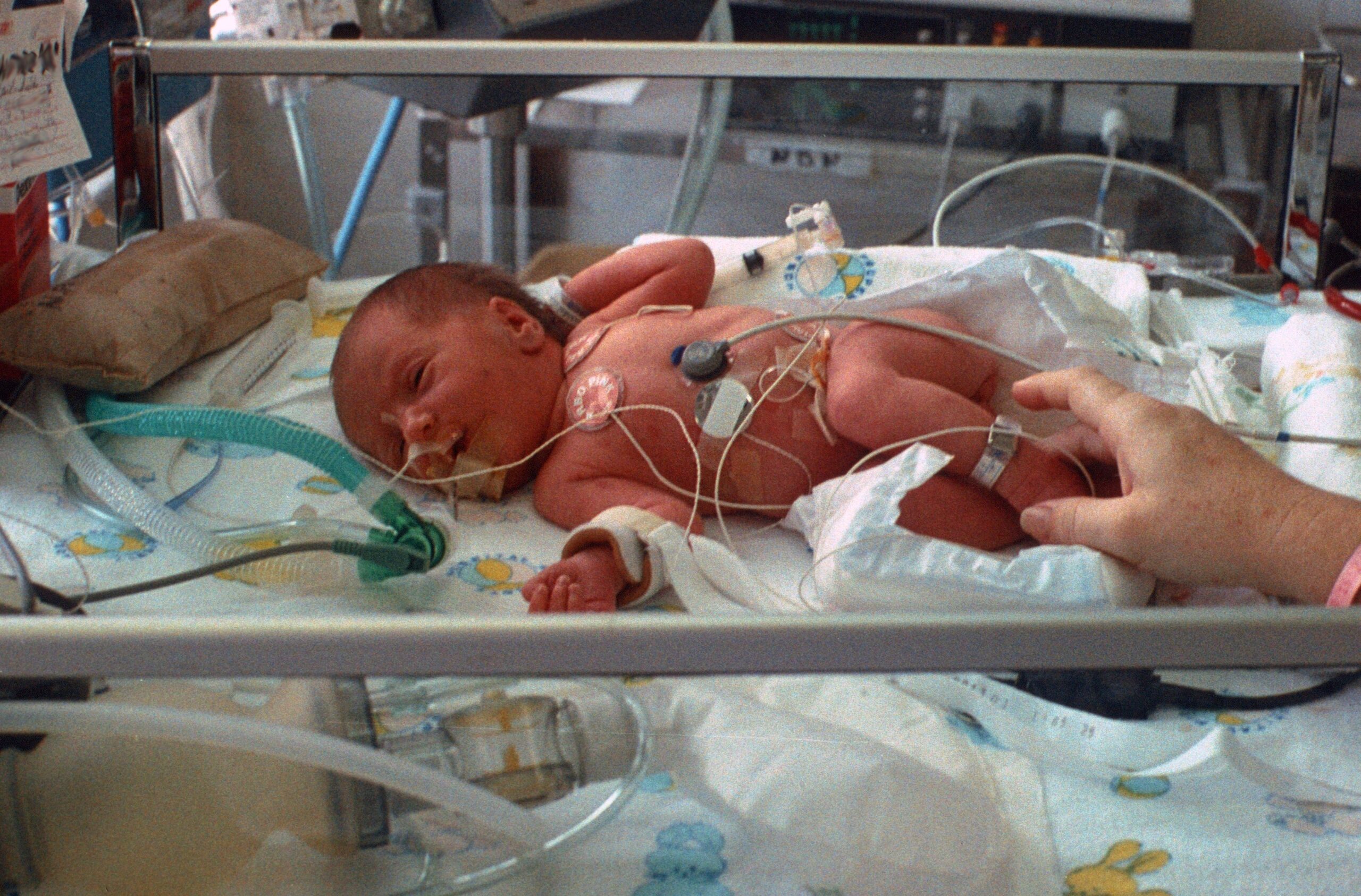
Periventricular leukomalacia (PVL) is a type of brain injury that primarily affects premature infants. It involves the death of small areas of brain tissue near the ventricles (the fluid-filled spaces in the brain), often due to insufficient blood flow or oxygen. The symptoms of PVL can vary depending on the severity and extent of the brain injury, but common symptoms include:
1. Motor Impairments:
- Spasticity: Increased muscle tone or stiffness, leading to abnormal movements or postures.
- Poor Muscle Control: Difficulty with coordination and fine motor skills.
- Weakness: Reduced strength or muscle tone, particularly in the arms or legs.
2. Developmental Delays:
- Motor Development: Delays in achieving milestones like sitting, crawling, or walking.
- Speech and Language: Delays or difficulties in speech and language development.
3. Cognitive Impairments:
- Learning Difficulties: Challenges with attention, memory, or processing information.
- Intellectual Disabilities: In severe cases, there may be significant cognitive impairments.
4. Sensory Issues:
- Visual Problems: Potential issues with vision, such as strabismus (crossed eyes) or other visual disturbances.
- Hearing Problems: Difficulties with hearing may occur, although they are less common.
5. Seizures:
- Types: Infants with PVL may experience seizures, which can range from mild to severe.
6. Motor Coordination Problems:
- Gait Abnormalities: Issues with walking or balance.
- Difficulty with Fine Motor Skills: Problems with tasks requiring precise hand movements, such as grasping objects.
7. Behavioral Issues:
- Hyperactivity or Attention Problems: Difficulty with attention and increased activity levels.
8. Neurological Signs:
- Abnormal Reflexes: Reflexes that are exaggerated or diminished.
- Tone Abnormalities: Issues with muscle tone, such as hypertonia (increased tone) or hypotonia (decreased tone).
9. Feeding Difficulties:
- Challenges: Problems with sucking, swallowing, or feeding can be present, especially in severe cases.
10. Visual or Cognitive Impairments:
- Potential Effects: Long-term visual or cognitive impairments may develop as the child grows.
Summary:
Periventricular leukomalacia often manifests with motor impairments, developmental delays, cognitive impairments, sensory issues, seizures, and other neurological signs. The severity of symptoms can vary widely based on the extent of brain injury. Early diagnosis and intervention are crucial for managing symptoms and improving outcomes for affected infants
What are the causes of periventricular leukomalacia?
Periventricular leukomalacia (PVL) is primarily caused by a reduction in blood flow or oxygen to the brain, particularly affecting the areas around the ventricles (fluid-filled spaces in the brain). The primary causes and risk factors include:
1. Premature Birth:
- Description: Infants born before 32 weeks of gestation are at higher risk for PVL.
- Mechanism: Premature infants have fragile blood vessels and less mature brain tissue, making them more susceptible to injury.
2. Intrauterine Infection:
- Description: Maternal infections, such as chorioamnionitis (infection of the amniotic sac) or other prenatal infections, can increase the risk of PVL.
- Mechanism: Infections can lead to inflammation and reduced blood flow to the brain.
3. Hypoxia-Ischemia:
- Description: A lack of oxygen (hypoxia) or reduced blood flow (ischemia) to the brain during or shortly after birth.
- Mechanism: Conditions such as umbilical cord complications, placental insufficiency, or birth asphyxia can contribute to hypoxia-ischemia.
4. Respiratory Distress Syndrome:
- Description: Common in premature infants due to underdeveloped lungs.
- Mechanism: Poor oxygenation and respiratory problems can lead to reduced oxygen supply to the brain.
5. Low Blood Pressure (Hypotension):
- Description: Low blood pressure in premature infants can reduce cerebral blood flow.
- Mechanism: Inadequate blood flow to the brain can result in ischemic injury.
6. Intracranial Hemorrhage:
- Description: Bleeding within the brain, particularly germinal matrix hemorrhage, can damage surrounding tissues.
- Mechanism: Hemorrhage can compromise blood flow and lead to ischemic damage, contributing to PVL.
7. Neonatal Cardiovascular Issues:
- Description: Conditions affecting the heart and circulation, such as patent ductus arteriosus (PDA) or congenital heart defects.
- Mechanism: Cardiovascular issues can affect blood flow and oxygen delivery to the brain.
8. Hyperbilirubinemia:
- Description: High levels of bilirubin in the blood (jaundice) in newborns.
- Mechanism: Severe jaundice can cause kernicterus, which may be associated with PVL.
9. Maternal Conditions:
- Description: Conditions such as preeclampsia (pregnancy-induced hypertension) can increase the risk.
- Mechanism: Preeclampsia can impair placental blood flow, leading to reduced oxygen and nutrients for the fetus.
10. Genetic and Environmental Factors:
- Description: Some genetic predispositions and environmental factors may contribute to the risk.
- Mechanism: Genetic factors may affect brain development and response to injury.
Summary:
The causes of periventricular leukomalacia are multifactorial, with premature birth being a primary risk factor. Other causes include intrauterine infections, hypoxia-ischemia, respiratory distress syndrome, low blood pressure, intracranial hemorrhage, neonatal cardiovascular issues, hyperbilirubinemia, maternal conditions, and potentially genetic or environmental factors. Addressing these risk factors through prenatal care and early medical intervention can help reduce the incidence and severity of PVL.
How is the diagnosis of periventricular leukomalacia made?
The diagnosis of periventricular leukomalacia (PVL) involves a combination of clinical evaluation, imaging studies, and sometimes laboratory tests. Here’s a detailed overview of how PVL is diagnosed:
1. Clinical Evaluation:
- Medical History:
- Prematurity: Assessing the infant’s gestational age and any complications during pregnancy or birth.
- Neurological Symptoms: Reviewing symptoms such as motor delays, abnormal muscle tone, or developmental delays.
- Physical Examination:
- Neurological Assessment: Evaluating motor function, muscle tone, reflexes, and developmental milestones.
- Developmental Screening: Identifying delays or abnormalities in motor and cognitive development.
2. Imaging Studies:
- Cranial Ultrasound:
- Purpose: This is often the first imaging test performed, especially in premature infants.
- Procedure: A non-invasive test that uses sound waves to produce images of the brain. It can identify areas of damage near the ventricles, where PVL occurs.
- Findings: May show areas of cystic changes or loss of brain tissue in the periventricular region.
- Magnetic Resonance Imaging (MRI):
- Purpose: Provides more detailed images than ultrasound and is often used to confirm the diagnosis or assess the extent of brain damage.
- Procedure: Uses magnetic fields and radio waves to create detailed images of the brain.
- Findings: Can reveal characteristic lesions associated with PVL, such as periventricular cysts and white matter damage.
- Computed Tomography (CT) Scan:
- Purpose: Less commonly used than MRI but can be helpful in some cases, especially if MRI is not available.
- Procedure: Uses X-rays to produce cross-sectional images of the brain.
- Findings: May show areas of cystic damage or calcification in the periventricular region.
3. Laboratory Tests:
- Blood Tests:
- Purpose: To identify underlying conditions or infections that could contribute to PVL.
- Tests: May include complete blood count (CBC), blood cultures, or tests for metabolic disorders.
4. Developmental and Neurological Assessment:
- Neurodevelopmental Evaluation:
- Purpose: To assess the impact of PVL on motor, cognitive, and behavioral development.
- Procedure: Includes standardized developmental tests and assessments by pediatric neurologists or developmental specialists.
5. Differential Diagnosis:
- Purpose: To rule out other conditions with similar symptoms.
- Conditions to Consider:
- Cerebral Palsy: A group of disorders affecting movement and muscle tone.
- Other Brain Injuries: Such as intraventricular hemorrhage or ischemic lesions.
Summary:
The diagnosis of periventricular leukomalacia is typically based on a combination of clinical evaluation, imaging studies (primarily cranial ultrasound and MRI), and sometimes laboratory tests. Early diagnosis through imaging, particularly in premature infants at risk, is crucial for managing PVL and planning appropriate interventions.
What is the treatment for periventricular leukomalacia?
The treatment for periventricular leukomalacia (PVL) focuses on managing symptoms, supporting developmental progress, and addressing complications. There is no cure for PVL itself, so treatment aims to improve quality of life and maximize the child’s functional abilities. Here’s a comprehensive approach to managing PVL:
1. Early Intervention:
- Developmental Therapies:
- Physical Therapy: Helps with motor skills, muscle strength, and coordination.
- Occupational Therapy: Assists with daily living skills and fine motor skills.
- Speech and Language Therapy: Supports communication and feeding difficulties.
2. Medical Management:
- Management of Neurological Symptoms:
- Medications: To manage symptoms such as muscle spasticity or seizures. Common medications include baclofen or antispasticity agents, and anticonvulsants for seizures.
- Regular Monitoring: Monitoring neurological development and adjusting treatments as needed.
3. Supportive Care:
- Nutritional Support:
- Feeding Assistance: May include feeding tubes or specialized diets if the child has difficulties with feeding or swallowing.
- Educational Support:
- Special Education Services: Tailored educational programs to address cognitive and developmental delays.
4. Management of Complications:
- Regular Monitoring:
- Vision and Hearing: Regular eye and ear exams to detect and address any sensory impairments.
- Orthopedic Issues: Monitoring and treating any musculoskeletal problems, such as scoliosis or contractures.
- Preventative Care:
- Vaccinations: Ensuring that the child is up-to-date with vaccinations to prevent infections.
5. Family Support and Counseling:
- Parental Support:
- Counseling and Support Groups: For parents to help manage the emotional and practical aspects of caring for a child with PVL.
- Educational Resources: Providing information on the condition and available resources.
6. Surgical Interventions:
- In Severe Cases:
- Orthopedic Surgery: For severe contractures or deformities that affect mobility.
- Other Surgeries: As needed for complications related to PVL.
7. Ongoing Follow-Up:
- Regular Check-Ups:
- Multidisciplinary Team: Ongoing care by a team of specialists, including neurologists, pediatricians, and developmental specialists, to monitor progress and adapt treatments.
Summary:
Treatment for periventricular leukomalacia involves a multidisciplinary approach to manage symptoms, support development, and address complications. Early intervention with therapies, medical management of symptoms, supportive care, and family support are key components. Regular monitoring and follow-up are essential to adapt treatment plans and optimize outcomes for affected children.
Leave a Reply
You must be logged in to post a comment.